Instruments can be reserved for use through the PPMS system. PPMS is an online accounting and equipment management software that can be accessed using your UK Login (linkblue). New users to the system must first request instrument access from within PPMS to make a reservation.
Light Microscopy Core Equipment
Equipment Reservation System
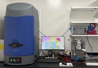
AMI HT Small Animal Imager
The Ami HT small animal imager from Spectral Instruments Imaging is capable of detecting both bioluminescence and fluorescence in vivo and in vitro. The system is available for imaging of immunocompetent rodents. It features 10 excitation wavelengths and allows imaging of up to 5 mice simultaneously. The integrated anesthesia system and mouse friendly platform enable easy and reliable imaging and analysis. The imaging acquisition/analysis software, Aura Imaging Software, provides advanced quantification and analysis of all data, and it is a free software available online for both PC and Mac users.
Key Imaging Techniques:
- Fluorescence imaging: works on the basis of the excitation of fluorochromes inside the subject by an external light source. This technique enables researchers to visualize and study various biological processes, including gene reporter analysis, fluorescence measurement of labeled molecule distributions, and so on.
- Bioluminescence imaging: based on light generated by chemiluminescent enzymatic reactions, particularly useful for luciferase-labeled models.
Nikon AXR Inverted Confocal Microscope
-
Special Features
- Heated stage inserts with CO2 and humidity for live cell work.
- Tokai Hit perfusion/media exchange system.
- Autosignal.ai.
- 25 mm field of view.
- Stage: Fully encoded scanning XY motorized stage for multipoint and tile scanning.
- Laser lines: multi-channel excitation with 405, 488, 561, and 640 nm lasers. Tunable emission bandwidth filters.
- Scanner: High speed resonant and galvanometric (8K scan area).
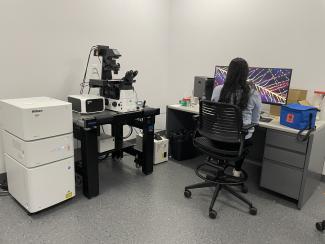
Nikon Super Resolution Inverted Microscope
-
Structured Illumination Microscopy (125 nm resolution)
- STORM Super Resolution (40 nm)
- Live cell imaging
- Total Internal Reflection Fluorescence Microscopy (TIRFM)
- DIC
- Time lapse imaging
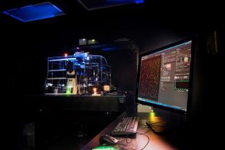
Nikon CSU-W1 SoRa Confocal Microscope
Spinning Disk Confocal with Super-Resolution Capabilities:
This system offers a unique combination of high-speed imaging and exceptional resolution without the need for specific sample preparation processes. Unlike standard confocal, which scans a single point at a time, the spinning disk confocal scans hundreds of points simultaneously. The addition of super-resolution imaging allows researchers to surpass the diffraction limit, achieving resolution down to 120 nm. What can be imaged in confocal mode can be further visualized in super-resolution, providing a seamless transition between imaging modalities.
Advantages of the spinning disk system include:
- Significantly faster imaging rates than standard confocal
- Superior temporal resolution with high-speed imaging with the ability to resolve biological processes not feasible with standard confocal.
- Improved Z-stack resolution for thick specimens, embryos, and organoids.
- Advanced live cell imaging capability including multiple particle tracking studies.
- Significantly reduced phototoxicity due to reduced light exposure
- Reduced photobleaching and improved imaging stability
- Ideal for tracking fast dynamics (ms regime) and extended timelapse (>24 hours)
Super resolution advantages:
- Resolution of 120 nm (XY)
- Realtime super resolution as the image is being collected
- Compatible with standard samples and fluorophores (no need for special dyes or sample conditions)
- Live Cell super resolution allowing fast dynamics to be captured at 100 nm resolution (no need to fix samples)
Hamamatsu ORCA-Quest 2 qCMOS camera:
- Ultra-low readout noise performance
- Suppress crosstalk and high QE
- High-speed readout
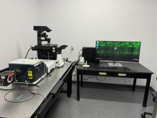
Zeiss LSM 880 Upright Multiphoton Microscope
-
Airyscan unit for super resolution
- Insight X3 extended wavelength laser
- High Sensitivity GaAsP Detectors
- In vivo imaging
- IACUC approved imaging facility
- Animal monitoring
- Isoflurane
- Multiphoton Imaging
- Imaging of cleared tissue
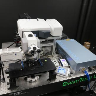
Nikon A1R Inverted Confocal Microscope
-
Galvano and Resonant Scanners
- Live Cell Imaging Incubator
- High Sensitivity GaAsP Detectors
- Confocal
- DIC
- Time lapse imaging
- Deconvolution
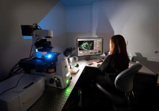
PicoQuant Fluorescence Lifetime Imaging (FLIM)
-
PicoQuant FLIM system is mounted on a Nikon A1 confocal microscope. It can be used to measure a fluorophore decay rate to detect molecular variations of fluorophores.
- PicoHarp 300 TCSPC module
- NKT SuperK High Power Supercontinuum White Light Laser
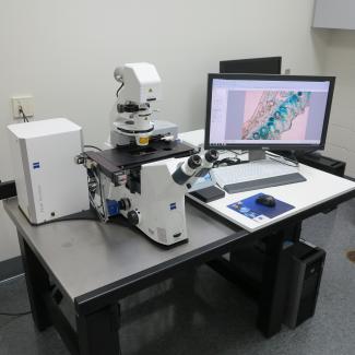
Zeiss Palm
-
Laser capture microdissection
- Colored camera
- Wide field Fluorescence
X-Clarity
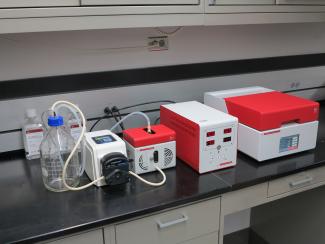
Tissue Clearing Services
The facility houses an X-Clarity tissue clearing system. The clearing process can be used to make tissue optically transparent. This allows for 3-dimensional imaging up to a few millimeters deep into tissues. To schedule a consultation, please fill out or contact form.
JPK Nanowizard 4 Atomic Force Microscope
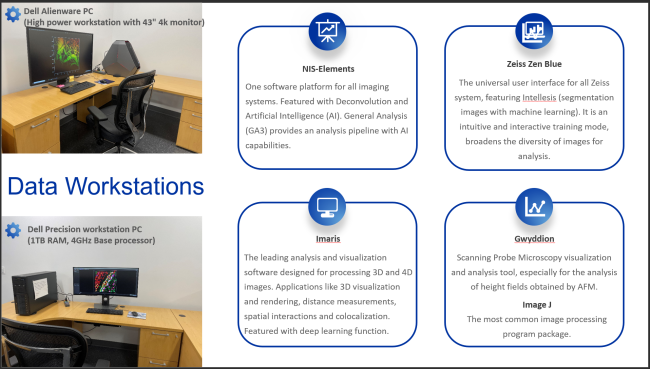
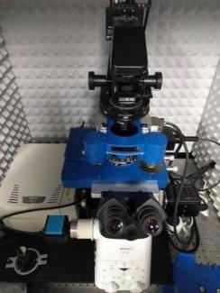
JPK Nanowizard 4 atomic force microscope mounted on a Nikon A1 confocal microscope. The hybrid scope allows the simultaneous and complementary investigation of samples in optical and nanomechanical/topographical ways. The Nanowizard combines fast tip scanning with high resolution scan seizes of up to 100 µm2. The unique architecture of the instrument, together with an acoustic enclosure and a stage top Petri dish heater, provides the mechanical and thermal stability to afford time laps imaging of living cells and observation of tissue dynamics in real time, while simultaneously covering the same optical and nanomechanical field of view. A comprehensive software suite facilitates many modes of data acquisition and analysis, including imaging force spectroscopy, direct overlay and nanolithography. Further, complementary and additional software for the analysis of AFM data, Gwyddion, is available on a separate workstation.
The Nikon A1 is equipped with four channels for confocal laser scanning and a high resolution water cooled sCMOS camera for fast and sensitive epi widefield fluorescence and brightfield/phase/DIC imaging.
Zeiss Axioscan Z1/ 7 Slide Scanners
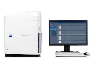
Both scanners allow automated scanning of up to 100 slides for either traditional histology stains or fluorescence labeled slides. The Z1 unit is also equipped with polarization function for bright field.
Standard Operating Procedures
User manuals and training documents for Light Microscopy Core equipment
Zeiss Palm Microcapture
PhysioSuite
Nikon SIM/STORM
Zeiss 880 NLO
Zeiss Axioscan Z1/Z7 Slide Scanner
JPK AFM/Nikon A1R Confocal
- Nanowizard 3 User Manual (pdf, 157 pgs)
X-Clarity
- X-CLARITY Tissue Clearing System Quick Start Guide (pdf, 8 pgs)
