Skip to main
University-wide Navigation

3T PRISMA Scanner

3T PRISMA Scanner

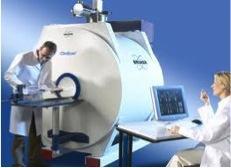
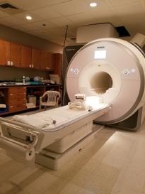
The UK-MRISC has a fully equipped 743 sq. ft. area for the whole-body Siemens PRISMA 3T MRI scanner. There are 64 and 20 channel head coils, 15 channel knee coil, spine array coil, two body matrix coils, and 3 flex coils. A state-of-the-art computer console running Syngo VE11C software as well as Syngo Via and Syngo Leonardo off-line workstations are available for examination and image processing using Siemens Software 3D rendering, image fusion, perfusion, DTI fiber tracking, spectroscopy, volume rendering, shaded surface display, multiplanar reconstruction, and maximum intensity projection. Supporting equipment includes an Avotec 6001 fMRI visual presentation system, MRA Inc and Cedrus-Lumina patient response systems, Redhat Linux analysis workstations, MRI compatible eye tracking system, Phillips CO2 monitor, and Biopac and Invivo physiological monitoring systems.

***RESEARCHERS - If you publish an article using the 3T Prisma scanner please be sure to cite the following grant in your acknowledgments/funding section: NIH/NIGMS S10OD023573***
 

7T BioSpec Scanner

7T BioSpec Scanner

The MRISC is equipped with a new 7T Bruker BioSpec, small animal, MRI scanner.

closed cycle, 14K cryocoil

This scanner is ideally suited for translational research with the capability to employ perfusion, diffusion tensor, anatomical, contrast, multinuclear spectroscopy, and functional techniques. We have experience imaging the heart, brain, spine, fat, lungs, vascular system, and cancer. 

An additional closed cycle, 14K cryocoil (above right image) provides a factor of a 2.5 to 5.3 increase in sensitivity for relevant applications.

SKYSCAN 1276 MicroCT

SKYSCAN 1276 MICROCT

A Skyscan MicroCT 1276 is available at the MRISC (Magnetic Resonance Imaging and Spectroscopy Center - https://www.research.uky.edu/magnetic-resonance-imaging-and-spectroscopy-center) in the basement of the Whitney-Hendrickson building. Usage training and a calendar reservation system are available. The 7T small animal MRI, a behavioral testing room, a necropsy room, and DLAR facilities are nearby.

Contact David K Powell, PhD (dkpowe2@uky.edu) for information about the Skyscan MicroCT.

MRISC SKYSCAN 1276 MICROCT Sample
Ramkumar T. Annamalai

The image to the right is from UK’s own Ramkumar T. Annamalai acquired on a similar system from his paper “Injectable osteogenic microtissues containing mesenchymal stromal cells conformally fill and repair critical-size defects” studying the repair of complex fractures with bone loss requiring a space-filling intervention to promote regeneration of bone.